貴金屬納米顆粒的表面等離子共振效應(yīng)[1]、尺寸效應(yīng)[2]、表面效應(yīng)[3]以及介電限域效應(yīng)[4]等因素使其具有獨特的光、電和催化等特性
因此,貴金屬納米顆粒在生物醫(yī)學檢測[5,6]、環(huán)境能源[7]、生物傳感[8]、光通訊[9]以及表面增強拉曼[10]等領(lǐng)域得到了廣泛的應(yīng)用
近年來,隨著納米技術(shù)的發(fā)展貴金屬納米顆粒的制備、特性和應(yīng)用成為相關(guān)領(lǐng)域的研究重點
因此,制備形貌可控、性能優(yōu)越的貴金屬納米顆粒極為重要
各向異性金納米棒,是近年來研究得較為深入的一種貴金屬納米顆粒
目前,制備金納米棒的主要方法有模板法[11]、光誘導法[12]、
電化學法[13]和種子生長法[14]
模板法使用合適的模板制備目標納米顆粒,但是產(chǎn)率較低且制備過程繁雜
光誘導法是在使用特定的反應(yīng)溶劑條件下對生長溶液進行不同波長的光輻射,但是生長的金納米棒形貌不均一且穩(wěn)定性較差
電化學法是對反應(yīng)溶液施加電場制備金納米棒,但是重復(fù)性不高、合成工序復(fù)雜且不可控
在種子生長法中顆粒的成核和生長分別進行,使產(chǎn)物的產(chǎn)率、形貌均勻性比較高[15]
使用種子生長法合成金納米棒,可通過改變生長液中各反應(yīng)參量調(diào)控其形貌和尺寸
鑒于此,本文研究生長液中AgNO3、CTAB、籽晶用量對金納米棒形貌及產(chǎn)率的影響,優(yōu)化反應(yīng)條件以合成形貌均一、產(chǎn)率高、分散性好、重復(fù)性高的金納米棒
1 實驗方法1.1 金種子的制備
用分析天平(型號為PX84ZH)稱取0.3553 g (0.1 mol/L)的十六烷基三甲基溴化銨(CTAB,分析純)溶于9.75 mL的純水中,然后將濃度為0.01 mol/L的氯酸金(HAuCl4·3H2O,分析純)溶液0.25 mL與上述溶液充分混合呈黃色溶液,在用磁力加熱攪拌器(型號為85-2型)劇烈攪拌下快速加入冰水現(xiàn)配的0.01 mol/L硼氫化鈉(NaBH4,分析純)溶液0.6 mL,溶液顏色由黃色變?yōu)椴枳厣砻鹘鸱N子形成,攪拌2 min后得到金種溶液,將其在室溫下靜置2-4 h得到金種子
金種子制備流程,如圖1所示
圖1
圖1制備晶種溶液的流程圖
Fig.1A Schematic diagram for the preparation of gold seed solution
1.2 金納米棒的合成
制備長徑比為3.8的小型金納米棒
用用分析天平稱取0.328 g (0.1 mol/L)的CTAB溶于9 mL的純水中,然后分別加入0.01 mol/L的HAuCl4溶液0.5 mL、0.01 mol/L的硝酸銀(AgNO3,分析純)溶液0.1 mL以及1 mol/L的HCl溶液0.2 mL,使其混合均勻后加入0.1 mol/L的抗壞血酸(AA,分析純)溶液0.08 mL,溶液的顏色由深黃色變?yōu)闊o色
用磁力加熱攪拌器攪拌2 min后快速加入1 mL種子溶液
將所得溶液在室溫下靜置過夜,制備過程如圖2所示
將最終得到的溶液用離心機(型號為HC-2518)以12000的轉(zhuǎn)速離心30 min,棄掉上清液后加入一定量的純水重新分散,離心分離兩次后得到金納米棒
圖2
圖2制備生長溶液的流程圖
Fig.2A schematic illustration of the synthesis of growth solution
1.3 金納米棒的表征和對福美雙的檢測
用分光光度計(型號為Lambda950)測試金納米棒的吸收光譜;用透射電子顯微鏡(型號為JEM2010)觀察金納米棒的形貌;用制得的金納米棒作為增強基底用于檢測農(nóng)藥殘留福美雙(Thiram,分析純)
用一次性針管吸取金納米棒溶液,滴一滴到面積為5 mm×5 mm的干凈玻璃片上,使其自然干燥
然后用分析天平稱取12 mg的福美雙粉末,用無水乙醇作溶劑配制成濃度為10-2 mol/L的福美雙溶液
然后用無水乙醇依次稀釋成濃度為10-3 mol/L-10-7 mol/L的待測溶液
最后將不同濃度的福美雙溶液分別滴到上述玻璃片上,待溶劑干燥后使用激光拉曼光譜儀(型號為HR Evolution)進行SERS檢測
拉曼測試的激發(fā)波長為785 nm,使用50倍物鏡,積分時間為10 s,掃描次數(shù)為2次,功率約為3 mW
2 結(jié)果和討論2.1 AgNO3 的用量對金納米棒的影響
種子生長法[16,17]:先制得小尺寸金納米顆粒作為籽晶,然后在有AgNO3、抗壞血酸和CTAB的混合溶液中加入一定量的籽晶,在籽晶的基礎(chǔ)上定向生長成金納米棒
調(diào)節(jié)AgNO3、籽晶和CTAB用量,可控制金納米棒的長徑比、形貌和大小
在合成金納米棒的過程中,AgNO3中的銀離子誘導金種向棒狀結(jié)構(gòu)演化,其用量對納米棒的長徑比和產(chǎn)率有很大的影響
圖3給出了分別加入0.035、0.065、0.08、0.1 mL的AgNO3 (0.01 mol/L),其余反應(yīng)物的濃度及用量不變時金納米棒的吸收光譜
從圖3可見,金納米棒的橫向共振吸收峰位于526 nm,縱向共振吸收峰分別出現(xiàn)在857 nm、848 nm、832 nm和813 nm
同時,隨著AgNO3用量的增加縱向共振吸收波長從857 nm藍移至813 nm
根據(jù)縱向共振吸收峰的峰位隨金納米棒長徑比的變化而變化可以推斷,金納米棒的長徑比隨著AgNO3用量的增加而減小
圖3
圖3AgNO3(0.035、0.065、0.08、0.1 mL)用量不同的金納米棒的吸收光譜
Fig.3Absorption spectra of gold nanorods prepared with different amounts (0.035、0.065、0.08、0.1 mL) of AgNO3
圖4給出了AgNO3加入量對金納米棒形貌的影響
可以看出,當加入0.035 mL的AgNO3時,金納米棒顆粒均勻性、分散性較好,產(chǎn)率較高(~96%),只有微量的球形顆粒,與圖3吸收光譜中對應(yīng)的高而窄的縱向共振吸收峰一致(圖4a)
加入0.065 mL的AgNO3時金納米棒的直徑幾乎沒有變化,只是兩端略微變短并出現(xiàn)微量的球形顆粒(圖4b);加入0.08 mL的AgNO3時金納米棒的分散性良好,只有少量的球形顆粒(圖4c);AgNO3用量增加到0.1 mL主要產(chǎn)物為金納米棒,有一些不規(guī)則的球形顆粒(圖4d)
這與圖3吸收光譜中縱向共振吸收峰分布較寬且強度不大一致
圖4
圖4AgNO3(a: 0.035 mL、b: 0.065 mL、c: 0.08 mL、d: 0.1 mL)用量不同的金納米棒的TEM照片
Fig.4TEM images of gold nanorods synthesized by different contents (a: 0.035 mL、b: 0.065 mL、c: 0.08 mL、d: 0.1 mL) of AgNO3 (Scale bars: 100 nm)
表1列出了AgNO3用量對金納米棒的平均長度、直徑、長徑比、縱向共振吸收峰以及產(chǎn)率的影響
可以看出,在其余反應(yīng)物用量及濃度保持不變的條件下,隨著AgNO3用量的增加金納米棒的平均長度和長徑比減小,縱向共振吸收峰波長從857 nm藍移至813 nm
4個樣品的數(shù)據(jù)表明,納米棒的產(chǎn)量較高(≥90%)
還可以看出,金納米棒的長徑比影響縱向共振吸收峰的位置,改變AgNO3用量可調(diào)控金納米棒的長徑比,從而調(diào)控其縱向共振吸收峰
金納米棒在近紅外區(qū)域具有可調(diào)諧的縱向共振吸收特性,使其在表面等離子共振領(lǐng)域有很好的應(yīng)用前景[18]
Table 1
表1
表1AgNO3用量對金納米棒平均長度、直徑、長徑比、縱向共振峰波長和產(chǎn)率的影響
Table 1Effect of AgNO3 contents on length, diameter, aspect ratio, longitudinal SPR and yield of gold nanorods
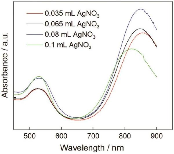
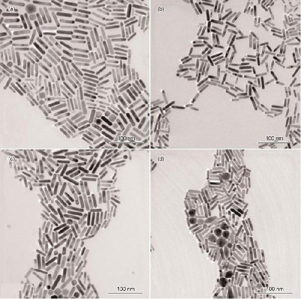
0.01 mol/L AgNO3 /mL
|
Length
/nm
|
Diameter
/nm
|
Aspect ratio
(R=L/D)
|
Longitudinal SPR
/nm
|
Yield
/%
|
0.035
|
44.5
|
10.5
|
4.2
|
857
|
96
|
0.065
|
39.1
|
9.6
|
4.1
|
848
|
98
|
0.08
|
38.2
|
9.7
|
3.9
|
832
|
94
|
0.1
|
32.9
|
8.9
|
3.7
|
813
|
90
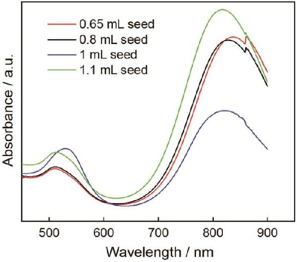
|
2.2 籽晶的影響
籽晶是金納米棒定向生長的“核”[19]
圖5給出了籽晶用量分別為0.65 mL、0.8 mL、1 mL和1.1 mL制備的金納米棒的吸收光譜
可以看出,籽晶量為0.65、0.8和1.1 mL制備的金納米棒,其吸收光譜中橫向共振吸收峰位置都約為511 nm,而縱向共振吸收峰位從855 nm減小至809 nm,峰的分布瘦而尖但是強度大
這表明,金納米棒的長徑比沒有明顯變化,主要產(chǎn)物是金納米棒
在籽晶用量為1 mL的吸收光譜中,出現(xiàn)位于530 nm的橫向共振吸收峰和位于814 nm處的縱向共振吸收峰,且縱向共振吸收峰峰型矮而寬
這表明,最終產(chǎn)物中有其它形貌的副產(chǎn)顆粒,金納米棒的產(chǎn)率不高
由此可見,只有籽晶的用量適當才能提高金納米棒的產(chǎn)率,減少副產(chǎn)顆粒
圖5
圖5籽晶(0.65 mL、0.8 mL、1 mL、1.1 mL)用量不同的金納米棒的吸收光譜
Fig.5Absorption spectra of gold nanorods prepared with different amounts (0.65 mL、0.8 mL、1 mL、1.1 mL) of seed
圖6給出了籽晶用量不同的金納米棒的TEM照片
從圖6可見,籽晶加入量為0.65 mL時的產(chǎn)物大多是金納米棒,尺寸分布較為均一,長度約為39.6 nm,只有微量的球形顆粒(圖6a)
籽晶加入量為0.8 mL時合成的金納米棒直徑較為一致,尺寸有長有短,形貌的均勻性略差(圖6b)
籽晶的用量為1 mL時產(chǎn)物中出現(xiàn)一些大小不一的球形顆粒,球形顆粒明顯增多,金納米棒的產(chǎn)率降低(~90%)(圖6c)
在圖5的吸收光譜中,位于820 nm的縱向共振吸收峰強度與其它譜線相差較大,且兩端分布較寬,從而證實了這一現(xiàn)象
繼續(xù)加大籽晶量至1.1 mL時金納米棒的產(chǎn)率明顯提高(~98%),且形貌均勻性、分散性更好,幾乎沒有副產(chǎn)顆粒(圖6d)
這與圖5吸收光譜中縱向共振吸收峰的中間細,兩端窄的情況一致
這表明,籽晶用量對金納米棒的形貌及產(chǎn)率有顯著的影響
生長液中籽晶不夠使金納米棒的產(chǎn)率降低;而籽晶用量過高則產(chǎn)物中出現(xiàn)副產(chǎn)顆粒
籽晶用量適當,有利于制備出高產(chǎn)率、高均一性的金納米棒
圖6
圖6籽晶(a: 0.65 mL、b: 0.8 mL、c: 1 mL、d: 1.1 mL)用量不同的金納米棒的TEM照片
Fig.6TEM images of gold nanorods synthesized by different contents (a: 0.65 mL、b: 0.8 mL、c: 1 mL、d: 1.1 mL) of seed (Scale bars: 100 nm)
根據(jù)不同籽晶量制備的金納米棒粒徑的分布,計算長徑比,結(jié)果列于表2
可以看出,當體系中其它反應(yīng)物用量和濃度一定時,金納米棒的長徑比與籽晶用量之間存在反比關(guān)系
隨著籽晶量的增加其長徑比明顯減小,縱向共振吸收峰波長發(fā)生藍移
這表明,對于一定數(shù)量的金原子,籽晶用量的增加使提供給每個籽晶的金原子數(shù)量減少,導致最終產(chǎn)物中出現(xiàn)副產(chǎn)顆粒
Table 2
表2
表2籽晶用量對金納米棒平均長度、直徑、長徑比、縱向共振峰波長和產(chǎn)率的影響
Table 2Effect of seeds contents on length, diameter, aspect ratio, longitudinal SPR and yield of gold nanorods
Au seed
/mL
|
Length
/nm
|
Diameter
/nm
|
Aspect ratio
(R=L/D)
|
Longitudinal SPR
/nm
|
Yield
/%
|
0.65
|
39.6
|
9.5
|
4.2
|
855
|
99
|
0.8
|
38.5
|
9.8
|
3.9
|
838
|
95
|
1
|
32.7
|
8.9
|
3.7
|
814
|
90
|
1.1
|
29.9
|
8.3
|
3.6
|
809
|
98
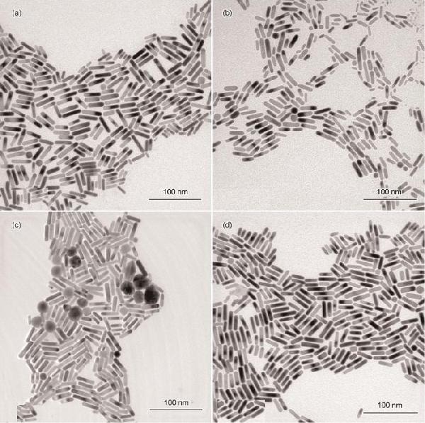
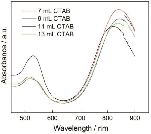
|
2.3 CTAB的影響
在金納米棒的合成過程中,表面活性劑CTAB以雙分子層形式擇優(yōu)吸附在金納米棒表面的不同晶面,從而控制金納米棒的生長[20]
圖7給出了用濃度為0.1 mol/L不同量的CTAB合成的金納米棒的吸收光譜
從圖7可以看出,4種金納米棒的吸收光譜十分相似,且橫向共振吸收峰位基本不變,而縱向共振吸收峰分別出現(xiàn)在815 nm、822 nm、837 nm及843 nm
峰值變化不明顯,說明金納米棒的長徑比幾乎保持一致
CTAB用量進一步增大至11和13 mL,縱向共振吸收峰發(fā)生變化,兩者均在859 nm處出現(xiàn)拐點
其原因是,CTAB用量過大使體系中Br?離子的強度提高
大量吸附在金納米棒表面的Br?離子抑制了金納米棒的定向生長,降低了金納米棒的產(chǎn)率,并產(chǎn)生副產(chǎn)顆粒[21]
圖7
圖7CTAB(7 mL、9 mL、11 mL、13 mL)用量不同的金納米棒的吸收光譜
Fig.7Absorption spectra of gold nanorods prepared with different amounts (7 mL、9 mL、11 mL、13 mL) of CTAB
在Au3+、AgNO3、籽晶和抗壞血酸的用量及濃度一定時,改變CTAB用量制備的金納米棒,其尺寸分布在圖8中給出
從圖8中的金納米棒透射電鏡照片可以看出,加入7 mL的CTAB時金納米棒的形貌均一性、分散性良好,且直徑較小,長徑比約為3.7,平均長度約為33.9 nm(圖8a)
這個結(jié)果,與圖7吸收光譜中位于841 nm處的縱向共振吸收峰強度高于其它曲線且峰型瘦而尖相對應(yīng)
加入9 mL的CTAB時產(chǎn)物中出現(xiàn)較多的不規(guī)則金納米顆粒,金納米棒的產(chǎn)率和形貌均勻性都不理想(圖8b)
這表明,生長液中過量的CTAB不利于金納米棒的生長
繼續(xù)增加CTAB用量至11 mL(圖8c)和13 mL(圖8d)時,產(chǎn)物大多為金納米棒和少量的大小不一的球形顆粒
金納米棒兩端類似于橢圓形,表面光滑和分散性良好,其平均長度約為35.5 nm和36.6 nm,長徑比大多為3.9
圖8
圖8CTAB(a: 7 mL、b: 9 mL、c: 11 mL、d: 13 mL)用量不同的金納米棒的TEM照片
Fig.8TEM images of gold nanorods synthesized by different contents (a: 7 mL、b: 9 mL、c: 11 mL、d: 13 mL) of CTAB (Scale bars: 100 nm)
使用粒徑計算軟件分析了不同CTAB用量的金納米棒的平均長度、直徑、長徑比、縱向共振吸收峰以及產(chǎn)率,結(jié)果列于表3
由表3可見,隨著CTAB用量的增加金納米棒的平均長度、直徑及縱向共振吸收峰波長僅發(fā)生了微小變化,其長徑比十分相近
這表明,在此反應(yīng)體系中下CTAB的加入量在一定范圍內(nèi)對合成的金納米棒的長徑比影響不大
但是從TEM圖可見,CTAB用量對產(chǎn)物的最終形貌及產(chǎn)率有較大影響
Table 3
表3
表3CTAB用量對金納米棒平均長度、直徑、長徑比、縱向共振峰波長和產(chǎn)率的影響
Table 3Effect of CTAB amounts on length, diameter, aspect ratio, longitudinal SPR and yield of gold nanorods
0.1 mol/L CTAB
/mL
|
Length
/nm
|
Diameter
/nm
|
Aspect
ratio
(R=L/D)
|
Longitudinal SPR
/nm
|
Yield
/%
|
7
|
33.9
|
9.2
|
3.7
|
815
|
96
|
9
|
32.7
|
8.6
|
3.8
|
822
|
87
|
11
|
35.5
|
9.1
|
3.9
|
837
|
89
|
13
|
36.6
|
9.3
|
3.9
|
843
|
93
|
2.4 在優(yōu)化條件下制備的金納米棒的光譜和形貌
在優(yōu)化條件下制備的金納米棒,其吸收光譜圖如圖9所示
在圖9中可見位于513 nm處的橫向共振吸收峰和位于817 nm處的縱向共振吸收峰,其中縱向共振吸收峰兩側(cè)分布較窄,強度高
圖10表明,金納米棒的形貌一致,分散性好,產(chǎn)率高,基本沒有副產(chǎn)顆粒
對金納米棒粒徑分布的計算結(jié)果,在圖11中給出
可以看出,金納米棒的長徑比約為3.8,平均長度約為34 nm
圖9
圖9在優(yōu)化條件下制備的金納米棒的吸收光譜
Fig.9Absorption spectrum of gold nanorods prepared under optimized conditions
圖10
圖10在優(yōu)化條件下制備的金納米棒的TEM照片
Fig.10TEM images of gold nanorods prepared under optimized conditions
圖11
圖11金納米棒的長度分布柱狀圖
Fig.11Length distribution histogram of gold nanorods
2.5 小型金納米棒對福美雙的拉曼光譜
圖12a給出了用金納米棒檢測不同濃度福美雙的拉曼增強光譜
可以看出,在金納米棒的增強譜線中均出現(xiàn)了明顯的福美雙特定的拉曼特征,分別位于552,1142,1375,1499 cm-1處
為了分析SERS強度與福美雙濃度的關(guān)系,以1374 cm-1處的特征峰強度為依據(jù),研究福美雙的濃度對SERS增強性能的影響
由圖12b可見,隨著福美雙的濃度由10-7 mol/L提高到10-2 mol/L,其特征峰的強度顯著提高
這表明,隨著福美雙濃度的提高金納米棒基底的SERS活性隨之提高,能檢測到的福美雙最低濃度達10-7 mol/L,低于規(guī)定的各食品中最大殘留限值
與其它功能化金納米棒基底對福美雙的SERS檢測體系相比[22,23],此法更加簡單可控且穩(wěn)定性高
圖12
圖12用金納米棒檢測福美雙的拉曼增強光譜以及1374 cm-1處的特征峰強度與福美雙濃度的關(guān)系
Fig.12Roman enhanced spectrum of different concentrations of thiram solution on gold nanorods substrate (a) and the relationship between the intensity of the 1374 cm-1 peak and thiram concentrations (b)
3 結(jié)論
(1) 用種子生長法可制備金納米棒
隨著AgNO3用量的增加Ag+引導金種定向生長成穩(wěn)定的棒狀結(jié)構(gòu),制備出的金納米棒形貌均一
籽晶的用量過少不能為金納米棒的生長提供充足的金種,使金納米棒的產(chǎn)率降低
CTAB用量過大則難以控制金納米棒不同晶面的生長速度,使金納米棒中出現(xiàn)較多不規(guī)則的球形顆粒
(2) 種子生長法的優(yōu)化條件:(0.01 mol/L) AgNO3用量為0.035 mL,(0.1 mol/L) CTAB用量為11 mL,籽晶用量為1.1 mL
在優(yōu)化條件下制備出的金納米棒形貌一致、分散性好,長徑比約為3.8,平均長度約為34 nm
(3) 這種小尺寸金納米棒具有較大的吸收截面和更高的光熱效率,可用于檢測低濃度生物分子
參考文獻
View Option 原文順序文獻年度倒序文中引用次數(shù)倒序被引期刊影響因子
[1]
Gu B Y.
Surface plasmon subwavelength optics:principles and novel effects
[J]. Phys, 2007, 36(04): 280
[本文引用: 1]
顧本源.
表面等離子體亞波長光學原理和新穎效應(yīng)
[J]. 物理, 2007(04): 280
[本文引用: 1]
[2]
Behafarid F, Matos J, Hong S, et al.
Structural and electronic properties of micellar Au nanoparticles: size and ligand effects
[J]. Acs Nano, 2014, 8(7): 6671
DOIPMID [本文引用: 1] " />
The effects of the dielectric environment on the optical extinction spectra of gold nanorods were quantitatively studied using individual bare and silica-coated nanorods. The dispersion and amplitude of their extinction cross-section, dominated by absorption for the investigated sizes, were measured using spatial modulation spectroscopy (SMS). The experimental results were compared to calculations from a numerical model that included environmental features present in the measurements and the morphology and size of the corresponding nanorods measured by transmission electron microscopy. The combination of these experimental and theoretical tools permits a detailed interpretation of the optical properties of the individual nanorods. The measured optical extinction spectra and the extinction cross-section amplitudes were well reproduced by the numerical model for silica-coated gold nanorods, for which the silica shell provides a controlled environment. In contrast, additional environmental factors had to be assumed in the model for bare nanorods, stressing the importance of controlling and characterizing the experimental conditions when measuring the optical response of bare surface-deposited single metal nanoparticles.
[5]
Ali M R, Wu Y, Tang Y, et al.
Targeting cancer cell integrins using gold nanorods in photothermal therapy inhibits migration through affecting cytoskeletal proteins
[J]. Proc. Natl. Acad. Sci. USA, 2017, 114(28): E5655
[本文引用: 1]
[6]
Nima Z A, Alwbari A M, Dantuluri V, et al.
Targeting nano drug delivery to cancer cells using tunable, multi-layer, silver-decorated gold nanorods
[J]. J. Appl. Toxicol., 2017, 37(12): 1370
DOIURL [本文引用: 1]
[7]
Bola?os-Benítez V, McDermott F, Gill L, et al.
Engineered silver nanoparticle (Ag-NP) behaviour in domestic on-site wastewater treatment plants and in sewage sludge amended-soils
[J]. Sci. Total Environ., 2020, 722(137794): 1
[本文引用: 1]
[8]
Paulo P M, Zijlstra P, Orrit M, et al.
Tip-specific functionalization of gold nanorods for plasmonic biosensing: effect of linker chain length
[J]. Langmuir, 2017, 33(26): 6503
DOIURL [本文引用: 1]
[9]
Sch?rner C, Adhikari S, Lippitz M.
A single-crystalline silver plasmonic circuit for visible quantum emitters
[J]. Nano Lett., 2019, 19(5): 3238
DOIURL [本文引用: 1]
[10]
Schlücker S.
Surface-enhanced raman spectroscopy: concepts and chemical applications
[J]. Angew. Chem. Int. Ed., 2014, 53(19): 4756
DOIURL [本文引用: 1]
[11]
Sanzortiz M N, Sentosun K, Bals S, et al.
Templated growth of surface enhanced raman scattering-active branched gold nanoparticles within radial mesoporous silica shells
[J]. ACS Nano, 2015, 9(10): 10489
DOIPMID [本文引用: 1] " />
A<strong> </strong>novel approach to high-aspect ratio gold nanorods has been presented. The gold nanorods as long as (200±18.62) nm with aspect ratio of above 10 were prepared with optimizing the concentration of surfactant cetyltrimethylammonium bromide (CTAB) by seed-mediated growth method at 25 ℃. Mechanism for gold nanorods formation is discussed. It is shown that the aspect ratio and longitudinal surface plasmon resonance (SPR) can be correlated with the concentration of CTAB. Moreover, by simply enhancing the ion strength of the reaction solution, the as-prepared gold nanorods can be purified for the different electrostatic aggregation effects between gold nanorods and spherical nanoparticles. Shape change of gold nanorods is confirmed by transmission electron microscopy images (TEM) and scan electron microscopy (SEM).
高 倩, 錢 勇, 夏 炎 等.
一種制備高長徑比金納米棒的新方法
[J]. 化學學報, 2011, 69(14): 1617
[本文引用: 1]
[22]
Pastorello M, Sigoli F A, Dos Santos D P, et al.
On the use of Au@Ag core-shell nanorods for SERS detection of thiram diluted solutions
[J]. Spectrochim. Acta A, 2020, 231(118113): 1
[本文引用: 1]
[23]
Yu Y, Zeng P, Yang C, et al.
Gold nanorod-coated capillaries for the SERS-based detection of thiram
[J]. Acs Appl. Nano Mater., 2019, 2(1): 598
DOI [本文引用: 1] class="outline_tb" " />
The effects of the dielectric environment on the optical extinction spectra of gold nanorods were quantitatively studied using individual bare and silica-coated nanorods. The dispersion and amplitude of their extinction cross-section, dominated by absorption for the investigated sizes, were measured using spatial modulation spectroscopy (SMS). The experimental results were compared to calculations from a numerical model that included environmental features present in the measurements and the morphology and size of the corresponding nanorods measured by transmission electron microscopy. The combination of these experimental and theoretical tools permits a detailed interpretation of the optical properties of the individual nanorods. The measured optical extinction spectra and the extinction cross-section amplitudes were well reproduced by the numerical model for silica-coated gold nanorods, for which the silica shell provides a controlled environment. In contrast, additional environmental factors had to be assumed in the model for bare nanorods, stressing the importance of controlling and characterizing the experimental conditions when measuring the optical response of bare surface-deposited single metal nanoparticles.
[5]
Ali M R, Wu Y, Tang Y, et al.
Targeting cancer cell integrins using gold nanorods in photothermal therapy inhibits migration through affecting cytoskeletal proteins
[J]. Proc. Natl. Acad. Sci. USA, 2017, 114(28): E5655
[6]
Nima Z A, Alwbari A M, Dantuluri V, et al.
Targeting nano drug delivery to cancer cells using tunable, multi-layer, silver-decorated gold nanorods
[J]. J. Appl. Toxicol., 2017, 37(12): 1370
[7]
Bola?os-Benítez V, McDermott F, Gill L, et al.
Engineered silver nanoparticle (Ag-NP) behaviour in domestic on-site wastewater treatment plants and in sewage sludge amended-soils
[J]. Sci. Total Environ., 2020, 722(137794): 1
[8]
Paulo P M, Zijlstra P, Orrit M, et al.
Tip-specific functionalization of gold nanorods for plasmonic biosensing: effect of linker chain length
[J]. Langmuir, 2017, 33(26): 6503
[9]
Sch?rner C, Adhikari S, Lippitz M.
A single-crystalline silver plasmonic circuit for visible quantum emitters
[J]. Nano Lett., 2019, 19(5): 3238
[10]
Schlücker S.
Surface-enhanced raman spectroscopy: concepts and chemical applications
[J]. Angew. Chem. Int. Ed., 2014, 53(19): 4756
[11]
Sanzortiz M N, Sentosun K, Bals S, et al.
Templated growth of surface enhanced raman scattering-active branched gold nanoparticles within radial mesoporous silica shells
[J]. ACS Nano, 2015, 9(10): 10489
PMID " />
A<strong> </strong>novel approach to high-aspect ratio gold nanorods has been presented. The gold nanorods as long as (200±18.62) nm with aspect ratio of above 10 were prepared with optimizing the concentration of surfactant cetyltrimethylammonium bromide (CTAB) by seed-mediated growth method at 25 ℃. Mechanism for gold nanorods formation is discussed. It is shown that the aspect ratio and longitudinal surface plasmon resonance (SPR) can be correlated with the concentration of CTAB. Moreover, by simply enhancing the ion strength of the reaction solution, the as-prepared gold nanorods can be purified for the different electrostatic aggregation effects between gold nanorods and spherical nanoparticles. Shape change of gold nanorods is confirmed by transmission electron microscopy images (TEM) and scan electron microscopy (SEM).
高 倩, 錢 勇, 夏 炎 等.
一種制備高長徑比金納米棒的新方法
[J]. 化學學報, 2011, 69(14): 1617
[22]
Pastorello M, Sigoli F A, Dos Santos D P, et al.
On the use of Au@Ag core-shell nanorods for SERS detection of thiram diluted solutions
[J]. Spectrochim. Acta A, 2020, 231(118113): 1
[23]
Yu Y, Zeng P, Yang C, et al.
Gold nanorod-coated capillaries for the SERS-based detection of thiram
[J]. Acs Appl. Nano Mater., 2019, 2(1): 598
Surface-enhanced Raman scattering (SERS)based capillary system is a promising route toward fast, real-time, and in-situ detection using a facile sampling process. Here, we demonstrate for the first time resonance-tunable SERS-active capillaries with high sensitivity, reproducibility, and stability. The strong signal consistency independent of measurement spots or storage time supports the long-term storage and signal tracking of analytes in practical use. The capillaries were successfully applied to the in-situ detection of pesticide residues, and the sampling process provides operation conveniency compared to conventional methods. These results indicate that our SERS-active capillaries have great potentials in fast in-situ detection for many practical applications.
表面等離子體亞波長光學原理和新穎效應(yīng)
1
2007
聲明:
“小型金納米棒的制備” 該技術(shù)專利(論文)所有權(quán)利歸屬于技術(shù)(論文)所有人。僅供學習研究,如用于商業(yè)用途,請聯(lián)系該技術(shù)所有人。
我是此專利(論文)的發(fā)明人(作者)